Our Services
Southern Heart provides a consultation service, and a full range of specialist cardiology tests and procedures.
What we do
Your GP will refer you to a cardiologist if you have a problem with your heart or if you're having symptoms that might be caused by your heart, like pain in your chest, dizziness or shortness of breath. The tests done at Southern Heart will help to work out what is happening with your heart – which diagnostic tests we do will depend on your symptoms. If you have been referred for a cardiology consultation, we will combine your clinical assessment and test results to come up with a comprehensive treatment plan.
How to prepare
The cardiology staff will inform you of the service you are to have and arrange for your procedure time.
Wear clothes and shoes suitable for exercising if you are booked in for an exercise ECG (stress test) or exercise stress echocardiogram, as you will be walking on a treadmill.
Bring your medication with you.
Unless we tell you otherwise, if you are having an exercise test please do not take any medication to lower your heart rate in the 24 hours before your test (for example Atenolol [Tenormin], Metoprolol [Betaloc, Lopressor], Diltiazem, Verapamil, Sotalol, Carvedilol [Dilatrend], Bisoprolol [Bosvate]), Digoxin.
You are welcome to bring a support person.
Please contact us if you have tested positive for Covid, or if you are feeling unwell.
Our consultation service
Our experienced cardiologists offer a consultation service to work with you to develop a management plan. This might be to reduce your overall risk, to work out the cause of any symptoms you have been having, or to treat a specific problem. We will discuss your cardiac symptoms, your general health, look at the medicines you are currently taking and any test results. We will put this information together with your preferences to recommend any further tests or treatments, including advice on changes to your lifestyle, such as exercise or diet, adjusting medications or considering procedures.
Our diagnostic tests

Resting ECG
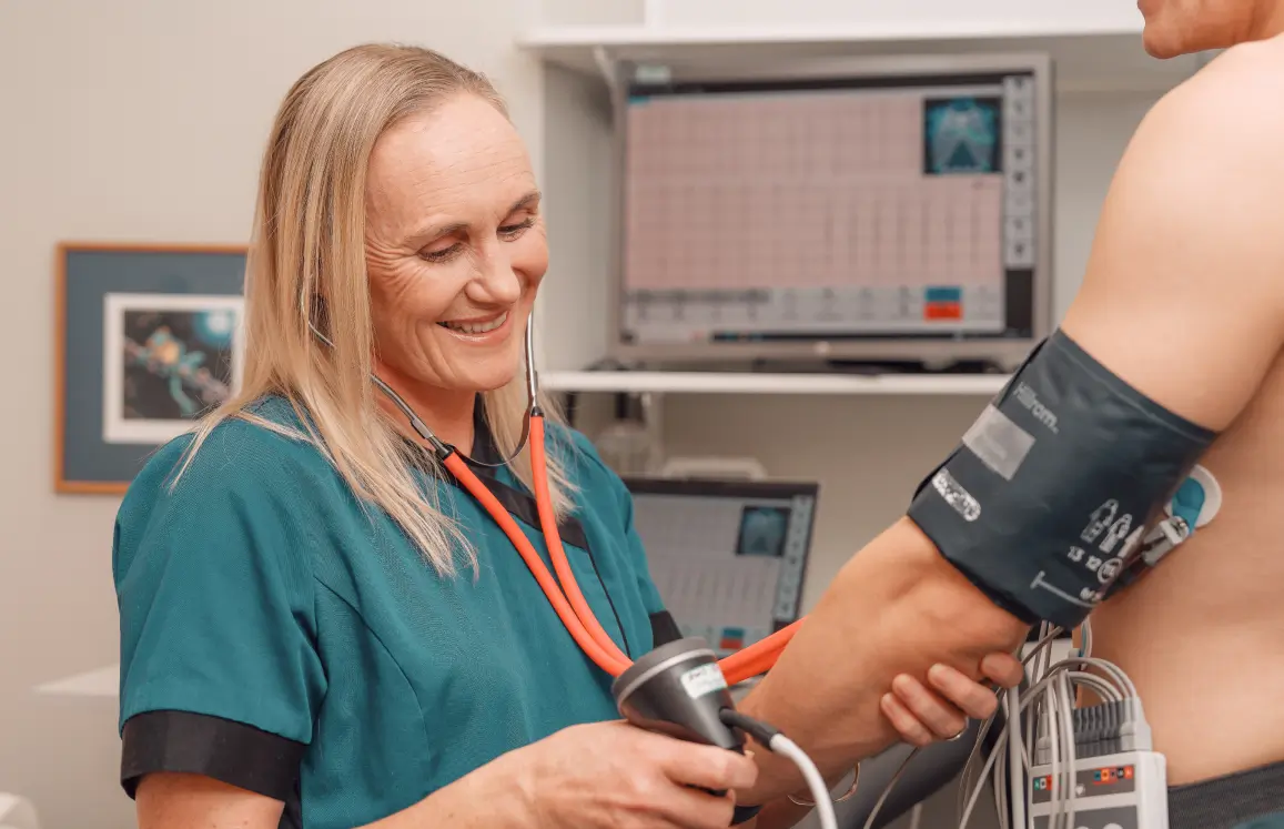
An electrocardiogram (ECG) machine records your heart's rhythm onto paper for us to analyse. The test is done using sticky electrodes placed on your chest, arms and legs. This helps us to detect abnormalities including problems with the conduction of the heart or heart rhythm abnormalities. This test usually takes five to 10 minutes.

Echocardiogram
An echocardiogram (Echo) test uses ultrasound to study the structure of your heart and how the heart and valves are working. A sensor is passed over your chest and heart which sends out and records these sound waves showing a moving image of your heart on a screen. This test takes about 30 minutes, then the images are analysed afterwards to produce a final report.

Exercise ECG
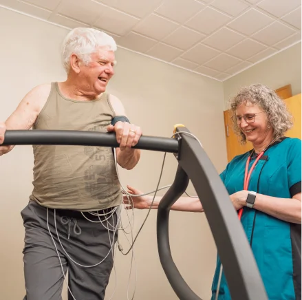
An exercise ECG (stress test) is used to show how your heart is coping when you are exercising, instead of resting. While you walk on a treadmill, which will slowly get faster and tilt up hill, your heart rhythm and blood pressure will be recorded. This test takes about 30 minutes.

Exercise stress echocardiogram
This test is a stress test with both ECG and echo. An echocardiogram is taken while you are at rest. You then exercise on the treadmill to show how your heart is coping when you are exercising, and another echo is done while your heart is beating fast. This test takes about one hour.

24 hour Ambulatory BP Monitor
A small blood pressure machine is attached to a belt around your body and is connected to a cuff around your upper arm. It measures your blood pressure at regular intervals over a 24-hour period – this is the most accurate way to get a true picture of what your blood pressure does over a full day, and can be used to make sure you don’t have high blood pressure just when you are seen by a doctor (white coat hypertension). It will also pick up times when the blood pressure is normal when measured in a clinic, but goes up at other times . You will be asked to keep a diary of what you do while wearing the monitor.

24 - 48 hour Holter monitor
The Holter monitor is a small, portable, battery powered ECG machine worn at home over a 24-hour period to assess the heart rhythm. It will record your heart rate and rhythm over this time, and you will be asked to keep a diary of what you do and any symptoms that you experience while you are wearing the Holter monitor. We will fit the monitor, show you how it works, and give you a courier bag to return it to us when the monitoring time is over. Once the monitor is returned, our physiologists and cardiologists will analyse the data and produce a report.

Event monitor
An event monitor is also used to assess the heart rhythm, and is typically worn by a patient for 5-7 days, depending on the frequency of the symptoms. You signal an "event" whenever your usual symptoms occur by pressing a button to activate the device. The heart rhythm is stored in the device memory. You will also be asked to keep a diary of what you do and any symptoms that you experience while wearing the monitor. We will give you a courier bag to send the monitor back to us when the monitoring period is over, and we will then analyse it.

Cardiac MRI
A cardiac MRI is an advanced cardiac imaging scan. We use it most commonly to look at the heart muscle in more detail, in particular looking for scarring or inflammation inside or around the heart. We also use cardiac MRI to look at heart valve disease, masses inside the heart, and the aorta. We will refer you to Pacific Radiology Group for this, with cardiology reports provided by Southern Cardiac Imaging. The scan takes 60 to 90 minutes.

Cardiac CT
CT calcium scoring is a simple CT scan that does not use X-ray dye, and helps provide a better risk assessment in people without any symptoms of heart disease. A CT coronary angiogram provides a direct non-invasive image of your heart arteries, and allows us to see if there is any build-up of cholesterol in the arteries, and, if there is, how much blockage there might be. We refer you for both of these types of cardiac CT to Pacific Radiology Group, with cardiology reports provided by Southern Cardiac Imaging. We may give you additional medicine to slow the heart down to obtain the best image, which you would take for a couple of days beforehand and on the day of the CT. This scan may take 30-90 minutes, depending on preparation time.
Our cardiology procedures
The cardiology procedures offered at Southern Heart provide minimally invasive therapies, and a much more detailed understanding of the heart.
Procedures:

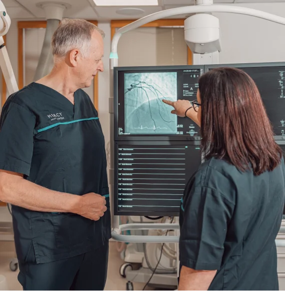
Coronary angiography
Coronary angiography is a test that examines the coronary arteries. Under local anaesthesia, a fine tube (catheter) is put into an artery of an arm or leg, passed into the arteries of the heart, and x-ray dye (contrast) is injected. The blood vessels are then x-rayed while the heart pumps. The resulting picture (angiogram) gives us the highest resolution imaging to see any problems with the arteries such as blockages.

Percutaneous coronary intervention (stenting)
Percutaneous coronary intervention (PCI), also called stenting, is a non-surgical procedure used to treat narrowing of the coronary arteries of the heart. Following an angiogram, a permanent metal scaffold known as a coronary stent is placed to hold narrowed coronary arteries open, restoring arterial blood flow to the heart muscle.
Transcatheter aortic valve implantation (TAVI)
TAVI is a procedure that treats a narrowed aortic valve (aortic stenosis) without requiring open-heart surgery. An artificial valve will be implanted into your heart. This is performed under a local anaesthetic and uses a tube inserted into a large blood vessel in the groin to deliver the valve to the heart.
Transcatheter Mitral valve edge to edge repair (TEER)
During this procedure a thin flexible tube, called a catheter is introduced into the body via a vein in your groin / upper leg. The device is then introduced into the body using this catheter and is positioned on the mitral valve holding or 'clipping' the leaflets together in a way that reduces the leakage.

Pacemaker insertion
A pacemaker provides additional heartbeats if your heart is too slow. It is a small box that usually is placed near the collarbone, with one or more wires going to the heart. The pacemaker listens to the heart, and, if you need it, generates an electrical pulse to start the heart beat. The insertion is done under local anaesthetic, with X-ray and ultrasound guidance to put the wires in the correct position.

Cardioversion
Cardioversion is a medical procedure to convert an abnormal heart rhythm into a normal rhythm using electricity. You will have a short general anaesthetic for this. While you are under anaesthetic, we will give you a therapeutic dose of electric current to the heart at a specific moment in the cardiac cycle. This “resets” the electrical activity of the heart, giving the normal heart rhythm a chance to re-establish itself.
Transoesophageal echocardiogram
Once in the oesophagus, this allows us to get highly detailed images of the heart and its internal structures, especially of the heart valves, and areas of the heart difficult to see from the outside.